Imaging and Wear Analysis Lab
Microscopes
- Olympus BX53M metallurgical microscopes with SC180 and DP74 cameras and Stream image analysis software
- Leica DM750P polarising microscopes with ICC50HD camera and Leica Application Suite imaging software
- Olympus IX73P2F inverted light microscope with Stream image analysis software
- A range of basic low power laboratory binocular microscopes
SEM
- Hitachi TM4000Plus Tabletop low-vacuum, 5-20kV SEM with Oxford Instruments EDS and AZtecOne analysis software
- Capable of low vacuum observation of samples up to 80mm in length without the need for sample coating. Magnification ranges from x30 to x10,000. The SEM is fitted with an Oxford Instruments EDS and AZtecOne analysis software, allowing chemical characterisation through point and area spectra, elemental mapping and line scans
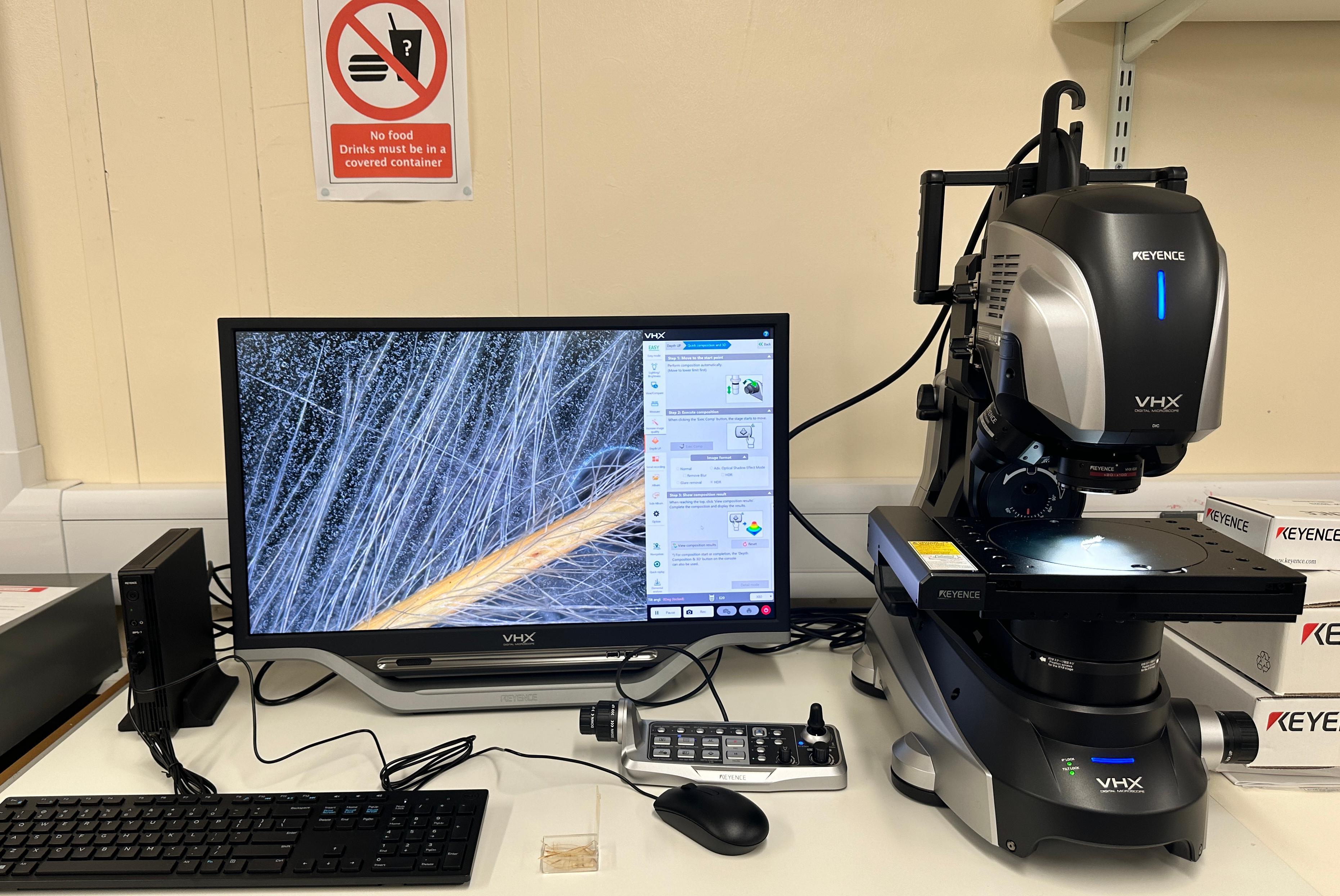
Keyence VHX-X1
The Keyence VHX-X1 offers:
- Advanced imaging and analytical capabilities able to reveal even the most subtle artefact features. The digital microscope offers a 300 mm stage suitable for artefacts to facilitate non-destructive analysis.
- A large depth of field with high resolution and a range of observation modes—including brightfield, darkfield, polarisation, and differential interference contrast. The Search Lighting function combines lighting from multiple directions enabling material observations previously undetectable using conventional optical microscopes.
- Elemental Analyser (EA) enhances usability by eliminating the need for vacuum environments, conductivity treatments, cutting, or other preparatory procedures. This streamlined process simplifies material identification and enables users to obtain instant results without compromising sample integrity.


